Ear - Anatomy and hearing
The sweetest of all sounds is praise.
Zenophon
Function
The ear is an organ able to receive sound. Sound we hear usually travels to the ear as vibrations of air molecules.
The ear is capable of detecting faint sounds and analyzing them, the direction they are coming from, the distance to the source of a sound, and the type of sound. This is done in the inner ear (cochlea) where the sound is changed to an electrochemical form that codes information about the intensity, frequency, and complexity of sound.
Another function of the ear is to keep the body balanced (in equilibrium). Equilibrium is related more to vision than hearing, but the sense organs for balance are found in the ear.
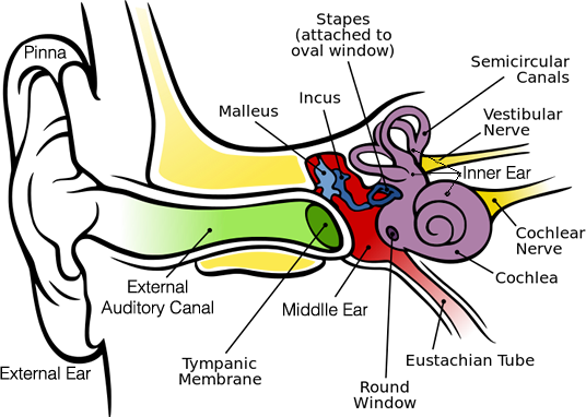
The ear's anatomy
Drawing adapted from Chittka L, Brockmann [ CC BY 2.5 ], Source Wikimedia Commons
The human ear can be divided into three areas: external, middle, and inner.
- External includes the part that sticks out from the head called the pinna. It also includes the hole entering the head called the auditory canal. It goes into the head until it comes to a dead end at the eardrum.
- Middle is a narrow air filled chamber separated from the outside by the eardrum ( tympanic membrane ) connected to a small chain of three bones called auditory ossicles . The three bones are the hammer ( malleus ), the anvil ( incus ) and the stirup ( stapes ).
- Inner is a complicated system of fluid filled passages and cavities deep in the temporal bone. It contains the sense organs for hearing, equilibrium, and the sensory nerve endings of the eighth cranial nerve.
Some important features.
Pinna
The pinna has no bones in it. A substance called cartilage is what makes it hard. The ear lobe is the only part of the pinna that has no cartilage.
The pinna has several small muscles connecting it to the skull and scalp. These usually do not function but some people are able to use them to wiggle their ears.
External auditory canal
The external auditory canal is the tube that ends at the eardrum (tympanic membrane). Its entire length is lined with skin, that also covers the eardrum (tympanic membrane). Fine hairs pointing out of the ear and a type of sweat gland that produces ear wax line it. Ear wax is secreted to discourage insects from entering the canal.
Eustachian tube
The main function of the eustachian tube is to keep the air pressure the same on both sides of the ear drum (tympanic membrane). The tube is closed during rest and opened during swallowing, so minor pressure differences are adjusted unconsciously. During a rapid dive in an airplane the tube may remain tightly closed. The pressure can be releaved by closing the mouth and nose and breathing out. This is called the Valsalva maneuver named after its discoverer Antonio Valsalva.
Otoscope
The instrument a doctor uses to look in your ear is an otoscope. She is looking at the color of the ear drum (tympanic membrane). The healthy membrane is pearl gray and sometimes has a pinkish or yellowish tinge. The doctor can also see through the membrane and the malleus is normally seen dividing the membrane into two equal halves. Sometimes the incus can be seen.
Cochlea
The cochlea is named after the Greek word for snail.